Mapping HSQC Chemical Shift Changes To Protein Structures
Mapping HSQC Chemical Shift Changes To Protein Structures
NOTE: You must first have your chemical shift assignments completed and a full structure or homology model before running this.
NOTE: For some homology models it will take more massaging of the “mapping_chem_shifts.awk” file for this to work; if there are any questions ask for help.
1.Open Linux or IRIX running environment
2.Move structure or homology model file (model.pdb) and “mapping_chem_shifts.awk” file into the same directory
3.Identify the chemical shift changes in your HSQC (NOTE: it helps to sort by residue number)
4.Open with vi editor the “mapping_chem_shifts.awk” file
5.In the “mapping_chem_shifts.awk” file you will see a list of if statements (see example). These are saying if the residue number in the pdb file matches the residue changing in the HSQC; insert a 10 to the 11 th column. For most .pdb files the residue number is in the 6 th column of the file. Append “mapping_chem_shifts.awk” so that all residues which correspond to a change in chemical shift ($6==37) add “10” to this 11th column or in other words residue 37 changed in the HSQC so it get a 10.
6.The VMD Explorer reads the numbering scheme as gradient of changes (10=change, 0=no change, or –10=opposite change). Therefore, there must be a -10 somewhere in the .pdb file. Find a residue distant from the bulk of changes and add the -10 to this residue ($6==120).
Example: Modifying mapping_chem_shifts.awk
if ($6==37) $11 =10.0
if ($6==45) $11 =10.0
if ($6==46) $11 =10.0
if ($6==66) $11 =10.0
if ($6==120)$11 =-10.00
7.After making the necessary modifications save and exit the awk script
8.Run “mapping_chem_shifts.awk” (awk –f mapping_chem_shifts.awk model.pdb >> out.pdb) (NOTE: this will run the awk script on your structure file and output a new pdb file.)
9.Open the newly created pdb file (out.pdb) with VMD Explore (NOTE: ask how to run VMD if you need help)
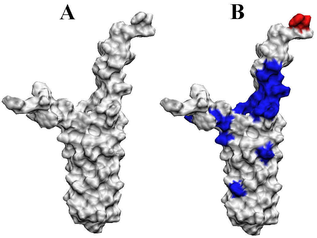
10.Generate a surface model or MS/MS model for the file and color by occupancy. For all residues labeled with a “10” in the new .pdb file (out.pdb) the color will change to blue. The blue regions of the protein indicate where the changes in the HSQC have taken place. (A=root protein structure B=mapped changes in HSQC mapped to protein surface blue=10 red=-10)
